Tuesday, May 26, 2015

Worm Blog Post

The earthworm is indigenous to Europe, but now can be found in Western Asia and North America. Earthworms live in moist soil, and don't have lungs or any other respiratory organs so they breathe through pores in their skin. Their skin must stay wet in order for oxygen to permeate their skin. Earthworms eat soil and decaying vegetation. A fun fact is that they have both male and female reproductive organs and reproduce by joining their clitellums together. Then they make a cocoon which holds the fertilized eggs together until they hatch.

Here is a link that full describes the dissection process:
http://youtu.be/m5P7s30NjxE


Sources:
 http://crescentok.com/staff/jaskew/isr/biology/biolab31b.htm

Frog Dissection Guide


The frog is found all over the world, but there are over 4,800 recorded species. Unlike most animals, the frog can breathe while submerged in water well as not submerged in water. When the frog is submerged the oxygen directly diffuses into the blood, but when they are not submerged in water it goes through as process called the Buccal Pumping. This Pumping system, pushes air through the throat and and breathes air through the nostrils. To eat, the frog uses their cleft tongue, instead of using hands or legs, to grab their prey. After they catch their prey, they use their eyes to help swallow their food and then down it goes down into their stomach. Generally it tends to eat many different types of insects. An interesting fact is that in Europe they tend to associate frogs with witchcraft while the Moche people of Peru believe that seeing a frog was good luck.


Here is a video that will help describe the dissection process of the frog:
http://youtu.be/L_OOOpvJbpg


Sources:
http://www.biologycorner.com/bio2/notes-frog.htmlthe frog

Grasshopper Dissection Blog

The grasshopper is a descendant of the cricket and there are over 11,000 species throughout the world of grasshoppers. Most grasshoppers are carnivores, but on occasion eat animal tissue and feces. In order for a grasshopper to eat, it uses its mandibles to chew its food and from there the salivary glands break down any carbohydrates. Eventually from there it goes through the oesophagus, the gizzards, and then finally the stomach where lots of enzymes begin to break things down. In order for the grasshopper to breathe they have an open circulatory system and their body cavities are filled with something called haemolymph which pumps fluid through the head and abdomen. For most grasshoppers that are bigger in size, they must keep moving in order to ventilate their bodies and help their abdominal muscles contract so they can breathe. An interesting fact is that if one has a grasshopper appear in a dream, some believe it is a sign of independence or a fear of "settling down". 


Here is a detailed description of how to dissect a grasshopper: 
http://youtu.be/SuGd0bb1vbM


Sources:


Crayfish Dissection Lab


The crayfish is known for living in freshwater areas, but are found in both the Southern and Northern Hemisphere. There are also many different species of crayfish throughout the world. When it comes to a crayfish's anatomy, they are very similar to a small lobster. The crayfish tend to breathe through their water-like gills, and they can survive both in and out of water for several days.  In order to eat, the crayfish use their claws to grab onto prey and then eat it with their mouth's. Usually crayfish tend to eat shrimp, water plants, insects, snails, and plankton. Crayfish are also known for being an Invasive species and also are consumed very often by people. 


Here is a lovely video describing the dissection of a crayfish:


Sources:
http://www2.sluh.org/bioweb/fieldbio/labsheets/invertebrateorganfunctions.pdf

Fish Dissection Blog


The Perch fish is found in Asia, Europe, Australia, and the United States and tends to live in small lakes, ponds, or rivers. Although it is found all over the world it is most commonly found in the Great Lakes in the U.S. In order to help this fish breathe, it has gills plus a lateral line system that helps detect vibrations in the water. Generally the perch eats shellfish, insect eggs, or other small fish that live in their habitat. An interesting fact about this fish is that there is a specific technique when one catches a perch, because it is commonly known for sallowing the hook. 


Here is a fantastic video describing the dissection of a Perch:
http://youtu.be/4dh3oWZIRCw


Sources: 
http://www.carolina.com/pdf/activities-articles/anatomy-perch.pdf

Starfish Dissection

There's are over 1,500 different species of starfish that live from anywhere from near the top of the ocean to the abyssal depths of the ocean. To help the starfish breathe, it has what is called the Hydraulic System which helps with gas exchange as well as moving and eating. This system is a series of canals that help transport different chemicals into the feet and lateral canals. In order to eat, the starfish takes in food from its mouth in the central disc and is moved into its gut (located towards the extends of the arms). From there it travels into many different tubes and canals and eventually the starfish will secret digestive enzymes which will absorb nutrients from the food. Generally the starfish tends to eat small fish, clams, oysters, and anthropods. The reason the starfish can eat animals that are larger then them Is because they are able to digest food outside their body. An interesting fact is that a starfish can produce both sexually and asexually and the oldest starfish was about 34 years old.


Here is a terrific video describing the dissection of a starfish (first link, ignore second link):
http://youtu.beSources: 
http://www.carolina.com/pdf/activities-articles/anatomy-starfish.pdf


Clam Dissection Blog


The clam is known to be found in both marine and freshwater habitats and each clam varies in both size and age. To breathe the clam uses gills located in the bivalve mantle cavity, and there is an open blood circulation system as well. This bivalve mantle cavity also helps a lot with with nutriention as well. To eat the clam collects food with their tentacles and then from their is transported to the mouth where it is filtered into respiration water. Generally clams like to eat larvae, detritus, eggs, and Protozoa. A fun fact about clams, is that for reproduction, both fertilization and larvae development happen in free water, rather than the clam itself. 

Here is a video describing the dissection of a clam:
http://youtu.be/Ebxhm6hA8io


Sources:
http://www.molluscs.at/bivalvia/
http://academic.evergreen.edu/t/thuesene/bivalves/Biology.htm

Thursday, March 5, 2015

DNA Restriction Lab

Purpose: The purpose of this lab was to not only understand how gel electrophoresis works,but also learn how to map restriction enzymes a DNA plasmid. Along with this, the lab was also supposed to help us learn about about DNA and how it is cut certain ways based on different variables.


Introduction:
Gel Electrophoresis often used in crime scenes and forscenics in order to compare two different sets of DNA. When the DNA is put into a gel electrophoresis, restriction enzymes are added to the a DNA sample so sciencetists are able to see where the those DNA fragments are compared to the DNA sample of either the criminal or victim. Restriction enzymes tend to cut up DNA into certain sections based on their nucleotide sequences and inside the cell they are usually made to break DNA Apart. In this situation, restriction enzymes are used to cut up a DNA sample to compare to another SNA sample. When adding the different restriction enzymes, you tend to add some idividually and some together with other restriction enzymes. Scientists do this because they want to find every possible way the DNA was spilt up so it can match a certain DNA fragment. 


Procedure:
We pipetted samples of DNA cut with various restriction enzymes into the grooves of a gel. We put the gel into a machine that runs an electric current through the gel, because DNA is negatively charged and will move towards the positive end. We stopped the electric current after 45 minutes to prevent DNA from running off the gel. Larger fragments do not travel as far in the gel because they cannot fit as easily through the pores in the gel.

Here is what the gel looked like before the electri current (sorry for the glare).

Here is what all the gels looked like after the electric current. Our gel is the middle gel in the bottom row.

After using the light and a marker, we made distinct lines of where the gels landed after the electric current.


Discussion: The results we got showed that restriction enzymes cut the DNA in a way so that many pieces of the DNA were the same as the model, showing that the negative DNA flowed properly from the negative pole of the gel to the positive side. In order to map the size of each fragment on the plasmid, we compare the length of the three lanes to the marker to understand roughly how many cuts and where the DNA underwent. Using PstI first, we saw that it had two fragments around 600 and 3300, and PSTI/ SSPI had three fragments at 600, 1500, and 1800. This means that the 600 length piece of DNA was never cut with the SSPI restriction enzyme, so it must have cut the longer piece of DNA into two, one slightly larger than the other. Similarly, the PSI/ HBAI gel cut the longer piece of DNA into two pieces, one much longer (at around 3000) and much shorter (around 300). Our gel was not as clear as other gels, so experimental error is at play in that certain lanes of the gel were not filled as much DNA as others. If all the lanes had an adequate amount of DNA, we might have been able to see our results more clearly. 


Conclusion: we successfully learned how restriction enzymes work and mapping the restriction enzymes on a piece of circular DNA. By loading gel and seeing where the restriction enzymes had cut, we can compare the effects of certain restriction enzymes and use them to compare the similarities in DNA. 

P-Glow E. Coli Lab


Introduction: The transformation of DNA is practice that many biologists use in order to have the bacteria expressed a trait it otherwise would not express. In order to do this, foreign DNA in a circular plasmid form must be added to the bacterial cell. Once there, the mechanisms of the cell are used to translate these genes into proteins that express the genes that were just added to the cell. The plates we are using gave agar, which is a nutrient rich medium that the bacteria thrives in under normal growing conditions. Ampicillin is an antibiotic that normally stops the bacterial cells from living and reducing properly. Arabinose is a sugar that bacteria use as a food source. 


Purpose: The purpose of this lab is to see if E. coli bacteria can successfully be transformed with a recombinant PGLO gene. If successful, the plates that were transformed will be antibiotic resistant as well as glow in the dark. 


Procedures: First we took two micro-test tubes, label them either +pGLO or -pGLO, added 250 micro-liters of transformation solution (CaCl2) to both containers, and placed the test tubes on ice. The using two different sterile loops, we added bacteria (E. Coli) to the loop and added it to the transformation solution to both micro-test tubes. After that, we took on sterile loop full of pGLO plasmid DNA (this glows a turquoise color when under a UV light) and added it to the +pGLO micro-test tube only. Then for ten minutes we let the micro-test tubes sit on the ice for ten minutes.

After the ten minutes we put both the +pGLO and -pGLO micro-test tubes in a water bath that was at 42 degrees Celcuis for exactly 50 seconds, and immediately put the micro-test tubes back on the ice.
After waiting two more minutes, we added LB nutrient brother to both of the micro-test tubes. After mixing the solution we put Exactly 100 micro-liters of either +pGLO to the plates labeled LB /amp and LB/amp/ara and -pGLO to the plates LB and LB/amp. (The LB means that any bacteria will grow, LB/amp means that only plasmid can grow, and LB/amp/ara means that arabinose can be used by pGLO genes to make a fluorescent protein) by using a sterile loop we spread the bacteria around the agar plate and then let the plates sit in an incubator (37 degrees C) for a day.
Adding the bacteria to the micro-test tube.

This is Kat adding the pGLO DNA plasmid to the +pGLO micro-test tube. This was very difficult to do.


The Colored containers in the turquoise Styrofoam are the micro-test tubes sitting on ice.
This is the micro-test tubes sitting in the cold water bath. 
This is spreading the bacteria on the agar plate.

Data:
Here is a data table of what each of the plates looked like after sitting in the incubator.


Here is a picture of a -pGLO LB/amp plate. There is no glow as well as there is no bacteria growth.

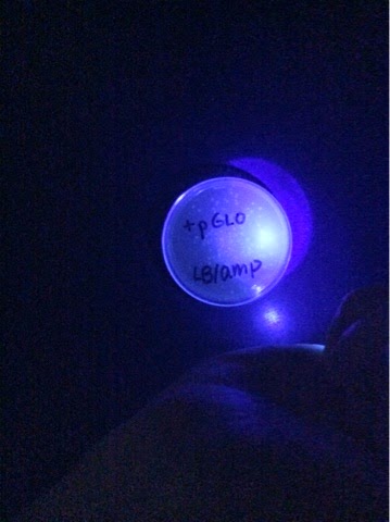
Here is a picture of the +pGLO LB/amp. There is no glow despite the fact that their was bacteria growth.
Here is a picture of the +pGLO LB/amp/ara plates. In this plate there is growth and it can glow under the UV light.

Analysis: Some observations we made about the amount of bacteria growth were that less bacteria grew on the plates containing ampicillin, despite the fact that we used the bacteria that contained the pGlo plasmid. Tis leads us to believe that not all of the bacteria were transformed, because there was much less growth compared to the control group of bacteria on the LB plate. We knew we were able to transform some of the bacteria, because there was no growth on the Amp plate when the bacteria did not have the plasmid, and when the bacteria did contain plasmid, there was some growth. Also, we knew they were successfully transformed because the bacteria did not glow in the dark when they did not have the plasmid.


Conclusion: We were able to successfully transform baceria using the pGlo plasmid, to make it resistant to ampicillin and to glow under UV light.

Monday, February 16, 2015

How to Extract DNA from a Strawberry


1. First we put the strawberry and a buffer in a plastic bag a squished it until it look like pulp (like the picture below).
2. Next we put the pulp into the funnel in order to only the liquid part of the pulp.
3. After there was enough of the liquid in the test tube, we put chilled alcohol into the test tube.

 4. Then, we let the alcohol sit for a couple minutes and eventually we could see the DNA floating around on top of the original liquid. The DNA looked very white and was just resting in the liquid. Below is a before and after picture of how the liquid looked before and after then alcohol had been in the liquid for awhile.


5. After the DNA began to show in the liquid, we were able to extract the DNA out of the liquid using a special tool. Below is a picture of the DNA we extracted.


Wednesday, January 7, 2015

Cell Communications Lab

Purpose:the purpose of this lab was to observe cell communication by measuring reproduction of yeast cells, which reproduce as a result of cell signaling. In this situation, the yeast signaled cells with the correct receptor protiens so the cells could reproduce with them. This was an example about how cell signaling is neccesary for the continuation of life.


Introducion: When only one type of yeast cell is present, either alpha-type or a-type, the yeast will reproduce asexually. However, in a mixed culture, the yeast will signal the other type with pheremones, which the other type have receptors for. This signals the yeast cells to grow towards each other, forming shmoos, and when they meet they reproduce. We measured the amount of cell communitcation happening by counting the shmoos in an area after a partictular amount of time.


Methods and Producers: 
In this experiment, we used had three different yeast samples: a-type, alpha-type, and mixed (a combination of a-type and alpha-type). After collecting each from an original culture, we put them in both agar plates and our own culture tubes. In the culture tubes, there was 2 mL of water along with a small clump of yeast. In the agar plates, there was 5 drops of yeast suspension (also know as yeast food). After doing both of this, we would take a forcep to put some of the yeast cells onto a coverslip, so we were able to see the different types of cells that were in the yeast. We first took a reading of the yeast immediately after putting the cells in the agar plates to find single haploid and budding haploid cells in the a- and alpha-types as well as single haploid and budding haploid cells, shmoos, and zygotes. After reading the yeast cells once, we put them into an incubator overnight so they yeast cells could mate and grow more. The next morning we again put bits of the yeast in a coverslip to see if there was difference in a moment of cells there were. When looking at cover slips, we also looked at many different fields of view in one coverslip, by changing where we were looking on the coverslip.


These are the agar dishes we put each of the yeast cells in to measure the different cells.


Here is the what the mixed culture looks inside of the microscope. Labeled is a budding haploid, and single haploid cell, a shmoo, and a budding zygote. 


After we finished the lab, we had to kill all of the yeast so it would not spread throughout the school. It was a sad ceremony, but they now live in peace.


The data for the a-type culture.


The alpha-type culture data.


The data for the mixed culture.

Discussion:
The data shows that shmoos would only form in the mixed agar plates and liquid culture, because alpha type and a type can sexually reproduce with one another. In general, the count for haploid, diploid, and shmoo for withon from the earlier time to the later time went up after incubating overnight. Since there were no shmoos forming in alpha or a type, alpha type yeast only has the receptor proteins needed for sexual reproduction and shmoo formation for a type, and vice versa. The incubation helps accelerates growth because the warmth was an ideal environment to grow. As tine passed, more signal could sent from one cell to another, pulling them together and causing the proliferation of yeast cells. Cell communication with pheromones There were problems with data collection and counting the number of yeast cells accurately, so if we were to do this again we would have made it more precise.  


Conclusion:
Because no shmoos formed in a non-mixed culture, the conclusion we can make is that the correct receptor proteins must be present, in order to recieve the signal and begin a response. An increase in time also increased the amount of reproduction occurring. Time allowed more signals to be sent, and more responses to be undergone.